Why the Clavicle Is the Most Underrated Structure in Male Aesthetics
In looksmaxxing discourse, most attention flows toward facial features — bone structure, skin quality, jawline definition. The frame receives less systematic treatment despite being the foundational variable that determines how every other physical attribute is perceived. A narrow frame with a perfect face reads differently than the same face on a broad, well-developed shoulder girdle. The clavicle is the structural reason why.
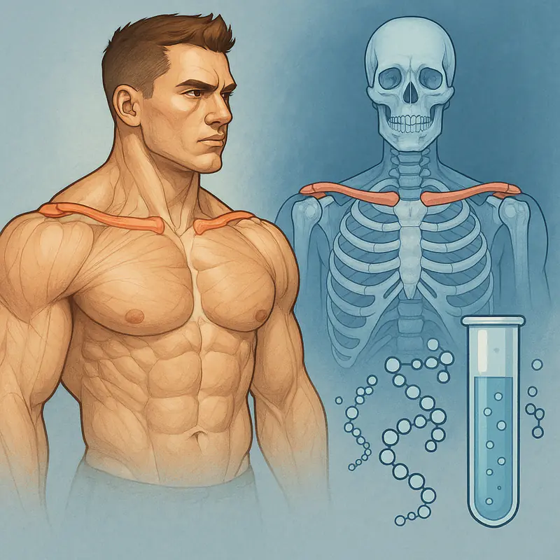
The clavicle — the collarbone — is a paired S-shaped long bone that spans from the sternum to the acromion of the scapula, forming the anterior strut of the shoulder girdle. Its lateral extent directly determines biacromial width: the distance between the outer edges of the shoulders. Biacromial width is one of the strongest single predictors of perceived male attractiveness in cross-cultural studies, with research consistently linking broader shoulders to higher ratings on dominance, health, and sexual attractiveness.
Unlike most long bones, the clavicle has a uniquely extended growth plate closure timeline. While the majority of long bones fuse their growth plates between ages 14–18, the medial epiphysis of the clavicle — the end that attaches to the sternum — is the last growth plate in the human body to close, typically between ages 22–28 and in some individuals as late as 30. This extended window has significant implications for researchers exploring structural optimization during early adulthood.
Bone Growth Biology: What Actually Drives Clavicular Development
Understanding which peptides are relevant to clavicular development requires understanding the biology of bone growth itself. Long bone growth occurs through endochondral ossification — a process by which cartilaginous templates at the growth plate (epiphyseal plate) are progressively mineralized and replaced by bone tissue. The rate and extent of this process is primarily regulated by three hormonal axes:
The GH/IGF-1 Axis: Growth hormone stimulates hepatic production of Insulin-like Growth Factor 1 (IGF-1), which directly drives chondrocyte proliferation in growth plates, osteoblast differentiation, and bone matrix synthesis. IGF-1 is the primary downstream mediator of skeletal growth — GH's effects on bone are largely IGF-1 dependent. The youthful GH secretion pattern — high-amplitude pulses, particularly during deep sleep — is what drives the rapid skeletal growth of adolescence. Its age-related decline is a central mechanism of the slower bone turnover and reduced structural development seen in adults.
Sex Hormones: Testosterone drives periosteal bone apposition — the widening of bones at their outer surface — and is responsible for the broader, denser skeletal architecture that distinguishes male from female frames. Estrogen paradoxically promotes growth plate fusion, which is why the testosterone-to-estrogen ratio during adolescence influences both the rate of bone growth and when it terminates. Males with lower estrogen conversion rates during puberty tend to have later growth plate fusion and thus a longer window for skeletal development.
Mechanical Loading: Bone is a mechanosensitive tissue. Osteocytes embedded in the bone matrix detect mechanical strain and signal osteoblasts to add bone in response to load. This is why resistance training during developmental years increases bone density and cross-sectional area — the mechanical signal is transduced into structural adaptation. The clavicle responds to the loading patterns of the muscles attached to it (primarily the pectoralis major, deltoid, and trapezius) and the compressive forces transmitted through the shoulder girdle during loaded exercise.
IGF-1 and Clavicular Growth: The Research Basis
IGF-1's role in skeletal development is among the most robustly established findings in endocrinology. Children with GH deficiency show dramatically reduced skeletal growth that is restored — including bone length and width — with GH replacement therapy, primarily through IGF-1 mediation. At the other end, acromegaly (pathological GH excess) produces excessive bone growth including clavicular widening, cranial changes, and hand and foot enlargement — confirming that the GH/IGF-1 axis actively drives adult bone remodeling even after growth plate fusion, primarily through periosteal expansion and bone density increases.
For looksmaxxing researchers, the relevant question is whether sub-pathological elevation of IGF-1 — within or near the upper range of normal physiological variation — produces meaningful structural differences. The epidemiological evidence is supportive: individuals with naturally higher IGF-1 levels (within normal range) consistently show greater bone mineral density, wider bone cross-sectional areas, and larger muscle mass compared to those with lower normal IGF-1, even controlling for body weight and training status.
Peptides that increase endogenous GH secretion — and consequently IGF-1 — therefore have mechanistic basis for influencing clavicular and shoulder girdle dimensions, particularly in individuals under 28 whose medial clavicular growth plates remain open, and for periosteal bone expansion in older individuals where growth plates have fused.
CJC-1295 and Ipamorelin: The GH Secretagogue Stack for Skeletal Development
The most extensively documented peptide approach to optimizing the GH/IGF-1 axis for structural development is the CJC-1295 + Ipamorelin combination. CJC-1295 is a GHRH analog that amplifies GH pulse amplitude throughout the day by extending the half-life of GHRH signaling. Ipamorelin is a selective ghrelin receptor agonist that generates clean GH pulses without the cortisol or prolactin elevation seen in older GHRP compounds.
Together, they recapitulate the youthful GH secretion pattern: sustained baseline amplification via CJC-1295 and sharp pre-sleep and pre-training pulses via Ipamorelin. The pre-sleep pulse is particularly relevant for bone remodeling — bone turnover is highest during the nocturnal GH secretory burst, and osteoblast activity is correlated with GH pulse amplitude.
Human studies confirm both compounds significantly increase circulating GH and downstream IGF-1. A 2006 study in Growth Hormone & IGF Research documented sustained GH elevation for 6 days after a single CJC-1295 dose. Ipamorelin's selectivity for GH release without cortisol activation is directly relevant for bone outcomes — cortisol is catabolic to bone, and GHRP compounds that elevate it alongside GH partially negate the anabolic skeletal signal.
For clavicular development specifically, the mechanistic logic is strongest in individuals with open medial growth plates (under ~25–28). In those with fused growth plates, CJC-1295/Ipamorelin's value shifts to periosteal bone expansion, increased bone mineral density, and the connective tissue and muscle development that directly influences the visual impact of shoulder width.
BPC-157: Connective Tissue Optimization for the Shoulder Girdle
The clavicle doesn't exist in isolation — its aesthetic contribution depends on the integrity and development of the surrounding connective tissue architecture: the acromioclavicular joint, sternoclavicular joint, coracoclavicular ligaments, and the tendons of the rotator cuff and surrounding musculature. These structures determine how well the shoulder girdle transmits force, maintains posture, and presents structurally under load.
BPC-157's dual angiogenic pathway (simultaneous VEGFR2-dependent and VEGFR2-independent vessel formation) drives healing and remodeling in tendons, ligaments, and joint capsules at a rate not seen with other research compounds. Published studies document significant improvements in torn tendon healing, ligament repair, and joint capsule integrity in animal models — with the mechanism centered on enhanced fibroblast migration, organized collagen deposition, and improved microvascularity in the repair tissue.
For looksmaxxers pursuing aggressive shoulder training to maximize the mechanical loading signal on clavicular bone and surrounding musculature, BPC-157's connective tissue protection is directly relevant. Shoulder injuries — particularly AC joint sprains, rotator cuff impingement, and biceps tendon issues — are among the most training-limiting injuries in heavy pressing and overhead work. Maintaining connective tissue integrity enables the consistent, progressive loading that drives both muscle development and the periosteal bone apposition that BPC-157's VEGFR mechanisms additionally support.
TB-500 (Thymosin Beta-4): Systemic Repair and Actin Regulation
Thymosin Beta-4 — and its research-accessible fragment TB-500 — is a 43-amino acid peptide that regulates actin polymerization throughout the body. Actin is the fundamental cytoskeletal protein in virtually all cell types, and its regulation by TB-4 governs cell migration, wound healing, cardiac muscle function, and tissue repair at a systemic level.
For structural development, TB-500's relevance is primarily indirect: it accelerates recovery from the tissue microtrauma that heavy training induces, reduces the inflammatory burden of high training volumes, and supports the satellite cell activity that drives muscle hypertrophy in trained tissue. Muscle development surrounding the clavicle — particularly the anterior and medial deltoid, upper trapezius, and pectoralis major — is what visually complements and fills out the skeletal frame. A wider clavicle without commensurate muscular development reads as underdeveloped; TB-500's recovery support enables higher training frequencies and volumes that drive this development.
TB-500 also promotes angiogenesis through a distinct mechanism from BPC-157 — upregulation of the Akt kinase pathway that drives endothelial cell migration and tube formation. Greater capillary density in trained muscle tissue improves both oxygen delivery during training and nutrient delivery during recovery — compounding the structural development response to progressive loading.
IGF-1 LR3: Direct Skeletal Growth Factor
IGF-1 LR3 is a synthetic analog of IGF-1 with an N-terminal extension that dramatically reduces binding to IGF-binding proteins (IGBPs) — the carrier proteins that limit IGF-1's bioavailability and half-life in circulation. Native IGF-1 has a half-life of approximately 10–12 minutes in free form; IGF-1 LR3's extended sequence increases this to approximately 20–30 hours while maintaining receptor affinity.
The downstream effect is sustained, elevated IGF-1 receptor activation across all tissues — including the growth plate chondrocytes in individuals with open epiphyses and the periosteal osteoblasts in those with fused plates. Research in animal models shows significantly enhanced bone growth rates with IGF-1 LR3 compared to native IGF-1 at equivalent doses, attributed to the reduced IGBP binding and prolonged bioavailability.
For clavicular development in individuals within the medial growth plate window (under ~28), IGF-1 LR3 represents a direct mechanism for stimulating endochondral ossification in the target structure. The medial clavicular epiphysis, as the last-fusing growth plate in the body, remains responsive to IGF-1 signaling longer than any other skeletal site — making it specifically relevant for researchers who have aged out of growth plate windows elsewhere but remain within the clavicular development timeline.
Compound purity and synthesis quality are paramount for IGF-1 LR3 research — the peptide's extended sequence makes it more synthesis-complex than shorter peptides, and impurities or sequence errors in lower-quality products produce neither valid research data nor reliable outcomes. Purity documentation, third-party testing comparisons, and supplier quality data for IGF-1 LR3 and the full range of structural development peptides are compiled at Peptides Clav, a reference used by the research community for sourcing evaluation across the clavicular and frame optimization peptide stack.
Mechanical Loading Protocols: Maximizing the Structural Signal
Peptides that optimize the GH/IGF-1 axis and connective tissue integrity are most effective when combined with loading protocols that provide the mechanical stimulus for bone adaptation. For clavicular and shoulder girdle development, the evidence points toward:
Overhead pressing movements (barbell overhead press, seated press variants) transmit compressive and bending loads through the acromioclavicular joint and along the clavicular shaft — the direct mechanical stimulus for periosteal apposition at the clavicle. Progressive overload in these movements over months produces measurable increases in bone density and cross-sectional area in loaded regions.
Lateral raise variations with controlled eccentrics load the deltoid-clavicle attachment and the ligamentous complex of the AC joint. High-volume lateral raises are a primary driver of medial and lateral deltoid development — the muscle bellies that visually widen the apparent shoulder width beyond skeletal width alone.
Farmer's carries and loaded hangs provide traction loading through the shoulder girdle, which some researchers propose may stimulate the sternoclavicular joint and medial epiphysis in individuals with open growth plates — analogous to spinal decompression loading. Controlled human data for this specific application is not published; the mechanism is mechanistically plausible but not directly confirmed.
The interaction between mechanical loading and the GH/IGF-1 axis is bidirectional: resistance training acutely stimulates GH release (and thus IGF-1), and elevated IGF-1 amplifies the bone's adaptive response to mechanical load. Peptides that chronically elevate GH/IGF-1 baseline therefore compound the response to training rather than substituting for it — making the combination of loading protocol and peptide stack mechanistically additive rather than redundant.
Protocol Design Considerations
A research protocol targeting clavicular and shoulder girdle development would logically integrate multiple complementary mechanisms: GH secretagogues (CJC-1295 + Ipamorelin) for hormonal axis optimization, BPC-157 for connective tissue integrity, TB-500 for systemic recovery and angiogenesis, and potentially IGF-1 LR3 for direct skeletal growth factor elevation in individuals within the clavicular growth plate window.
Timing considerations: CJC-1295 + Ipamorelin pre-sleep to leverage nocturnal GH secretion for bone remodeling; BPC-157 post-training for connective tissue repair in the acute recovery window; TB-500 2–3 times weekly for sustained systemic repair support. IGF-1 LR3, given its 20–30 hour activity window, is typically used on training days only to maintain IGF-1 receptor sensitivity.
All compounds referenced here are research chemicals with no FDA-approved therapeutic indications for aesthetic or structural development purposes. None should be approached without appropriate medical supervision, baseline hormone panel evaluation, and realistic assessment of the evidence gap between animal model data and confirmed human structural outcomes.
Conclusion: The Frame Is a Research Target
The clavicle's extended growth plate timeline — uniquely persisting to age 28 in many individuals — makes it the most accessible skeletal structure for researchers pursuing frame optimization during early adulthood. The GH/IGF-1 axis is the primary biological driver of clavicular development; peptides that optimize this axis have mechanistic basis for influencing outcomes during the open growth plate window and for periosteal bone expansion beyond it. Connective tissue peptides like BPC-157 and TB-500 support the training volumes necessary to provide the mechanical loading signal that bone adaptation requires.
The frame optimization approach — systematic, evidence-grounded, multi-mechanism — represents the same methodological rigor that effective looksmaxxing applies to facial aesthetics, skin quality, and body composition. The clavicle is not a fixed parameter. It is a biological structure subject to the same research-based optimization that governs every other dimension of appearance.